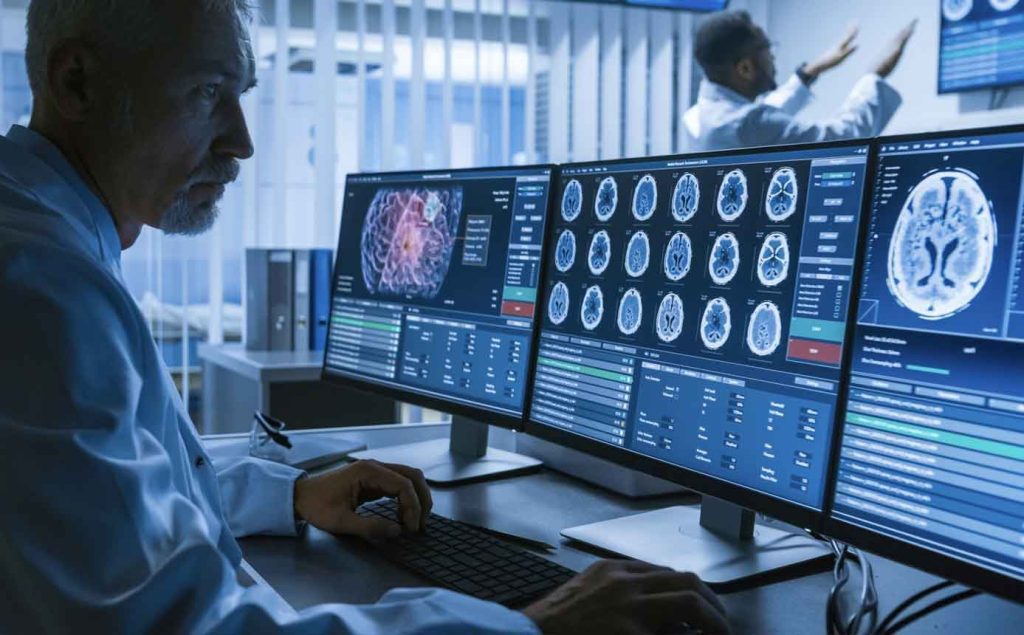
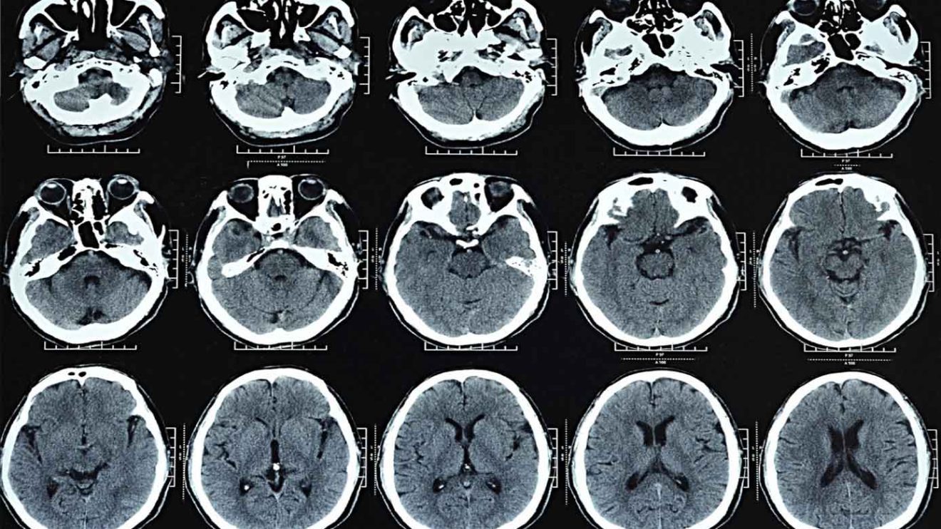
What is a head CT scan of the brain? When and how do you do it? What can she teach us? Does it have side effects? Instructions
CT stands for Computerized Tomography. This is a non-invasive test that is done using X-rays from several sources at the same time. The rays pass through the target organs (where you want to observe) and are picked up by the sensors.
The information captured by the sensors is processed in a computer, and it creates a high-quality, detailed, and accurate 3D image of the target organ. In our case, it is the head in general and the brain in particular.
How is the CT scan of the brain done?
The subject lies down on the examination bed, which then moves into the scanning machine (which looks like a large ring). During the scanning operation, the scanner moves in a circle around the patient’s head. As long as the scanner is running, lie still.
During the test, which usually lasts only a few minutes, the subject does not feel anything. There is no need for any anesthesia or sedation before the test.
Before the examination, the X-ray technician asks the subject to remove jewelry and metal objects because these may interfere with receiving a clear image.
What is the difference between a CT scan with and without contrast material?
In some cases, the attending physician will recommend a CT scan of the head using contrast material (a solution containing iodine). In such a case, the patient may be asked to come to the examination while fasting and to do blood tests in preparation for it. Specific instructions are given when the appointment is made.
Before the CT examination, an infusion (thin tube) is set up in the vein, and it is used to inject the contrast material during the examination. When the contrast material is injected, you sometimes feel a momentary feeling of heat and a metallic taste in your mouth.
3 different tests include the injection of contrast material:
- Head CT with contrast material is designed to emphasize differences between different tissues in the brain and helps to diagnose different pathologies (diseases).
- CT of the arteries – CTA (abbreviation of CT angiography) – makes it possible to get an image of the arteries in the brain and to see various damages caused to them such as blockage, abnormal expansion, or rupture.
- CT of the veins – CTV (abbreviation of CT venography) – makes it possible to get a picture of the cerebral veins and identify different pathologies such as blockages.
When is a head CT done?

Head CT is used for the following purposes:
- Imaging of the brain structure, the bones of the skull, and the bones of the face.
- Identification and location of processes within the brain tissue such as a tumor, bleeding, infection (abscess), diagnosis of stroke, diagnosis of birth defects, and accumulation of fluid in the brain.
- Identification of processes outside the brain tissue such as inflammations, fractures, infections, and tumors in the eye sockets or sinuses.
- In case of contusion, the test is done to rule out brain bleeding or bone fracture.
- Detection of damage to blood vessels in the brain (both arteries and veins).
What is MRI vs CT scan?
Both MRI vs CT scan are valuable tools in medical imaging, each with its unique strengths and applications. MRI excels at providing detailed soft tissue images and is particularly beneficial for diagnosing abnormalities in organs and soft tissues. On the other hand, CT scans are excellent for rapidly capturing images of bones, internal organs, and the skeletal system.
The choice between MRI vs CT scan depends on the specific clinical scenario, the area of the body being imaged, and the information required by healthcare providers to make accurate diagnoses and treatment decisions.
What are the possible risks and complications of a head CT scan?
This is a non-invasive, short, and painless test, and in most cases, you can return to normal activity immediately after it is finished. However, there are some important things to know about the test:
Radiation exposure During a CT scan, you are exposed for a short time to X-ray radiation. The level of radiation emitted by the new devices is not high and is even relatively low compared to other CT tests. The risk becomes significant only if a large number of tests are done.
Usually, the benefit that can be derived from the test exceeds the risk involved.
- CT scan of the brain during pregnancy. A woman who is pregnant or there is a possibility that she is pregnant should report this to the attending physician before doing the test. A CT scan is not recommended for pregnant women due to the danger that the radiation will harm the fetus. Breastfeeding women should avoid breastfeeding for 24 hours after the injection of contrast material.
- Reaction to the contrast agent (iodine). Rarely, an allergic reaction to iodine may develop. Allergic reactions are usually mild, but in rare cases, the reaction may be severe. That is why it is important to inform the doctor or the x-ray technician if you know of sensitivity to iodine.
- Also, a patient must inform the doctor or the x-ray technician if he knows that he suffers from one or more of the following diseases: asthma, diabetes, kidney disease, heart disease, and thyroid disease.
How long does a ct scan of the brain take?
The duration of a CT scan of the brain can vary depending on several factors, including the specific area of the body being scanned, the complexity of the examination, and whether contrast dye is used. In general, a CT scan typically takes anywhere from 10 to 30 minutes to complete. However, for more extensive scans or when the contrast dye is administered, the procedure may take up to 45 minutes to an hour.
What to do when the CT scan of the brain results are received?
A doctor specializing in imaging tests (radiologist) deciphers the test. You should go with the results of the test to the attending physician who gave the referral to perform the test.


Add Comment